Section: New Results
Classification of neurons to study Parkinson’s disease
Participants : Alexis Zubiolo, Eric Debreuve, Xavier Descombes.
This work has been made in collaboration with Michèle Studer's team at iBV
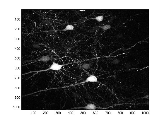
In this project, the goal is to perform unsupervised classification of rat neurons in order to study the Parkinson’s disease. The Institut de Biologie Valrose (iBV) provided us with 3-D images of rat cortices obtained by confocal microscopy. The discriminant features between normal and pathological neurons include the number of dendrites, the length and diameter of the apical dendrite, the shape and size of the soma For each neuron, these features have to be computed automatically from the images. The specificity of this problem is that, for each rat cortex, we are given several images:
-
one low resolution (LR) image which shows an overall view of the cortex and allows to compute the features related to the apical dendrite;
-
some high resolution (HR) images (typically between 4 and 6) which provide close-ups of the somas of the neurons and allow to compute the other features.
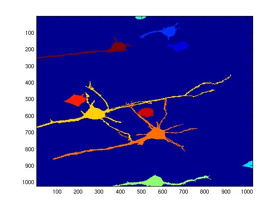
This work consists in (1) extracting the neurons from the images (see Fig. 12 ), (2) matching the corresponding neurons in the HR and the LR images, (3) computing the features for each neuron, and (4) classifying the neurons, for example using a kernel Support Vector Machine (SVM).