Section: Partnerships and Cooperations
Regional Initiatives
-
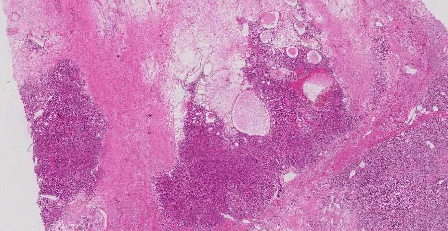
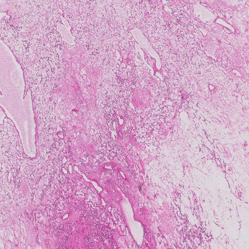
We started a collaboration with the team TIRO (Transporteurs en Imagerie et Radiothérapie en Oncologie), CEA/UNS/Centre Antoine Lacassagne, Nice, concerning the detection of tumorous cells in kidney histopathology (see Fig. 17 ). Although the images have a very high resolution, the problem is extremely difficult due to the similarity between different type of cells.
A coarse-to-fine approach seems perfectly adapted since the acquisitions are performed at several resolutions. Typically, six resolutions are available (see Table 1 ). However, contrarily to what is usually done, we do not plan to develop a unique approach, to apply it to the coarser resolution, and to use the corresponding result projected onto the following resolution as the initialization of the next step. Our idea is to think of which approach to take at each resolution level, and to gradually improve the detection confidence from “this broad area might contain tumorous cells” to “with high confidence, this small, finely delineated region is a tumorous cell”. For example, we might start with histogram analysis or simple thresholding methods on the coarser resolution. Then, texture analysis could be performed in intermediate resolutions. Finally, fine radiometric and shape analyses could be done on the full resolution image to achieve object-level detection.
-
We have a collaboration with the Laboratoire d'Océanographie de Villefranche (LOV), CNRS/Université Pierre et Marie Curie, concerning automatic classification of zooplankton organisms for an embedded system called UVP for Underwater Vision Profiler (see Section 6.12 ).
-
We have a collaboration with IPMC (H. Barelli) on vesicules tracking for characterizing cell membrane properties (see Section 6.7 ).